Fossils
Type of resources
Available actions
Topics
Keywords
Contact for the resource
Provided by
Years
Formats
Representation types
Update frequencies
Scale
-
Computed tomography (CT) scans of extant and fossil chondrichthyans, of teeth organized into functional dentitions. These scans were taken on a Nikon Metrology HMX ST 225, in the Image and Analysis Centre, Natural History Museum, London.
-
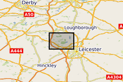
Reflectance Transformation Imaging files for the type specimen GSM105875 [IGSN:UKBGSGSM105875] and two paratypes GSM106040 [IGSN:UKBGSGSM106040] , GSM106112 [IGSN:UKBGSGSM106112] of Hylaecullulus fordi, a new species of rangeomorph from the Bradgate Formation (Ediacaran) of Charnwood Forest, UK. Supporting information for Kenchington, Dunn and Wilby - Modularity and overcompensatory growth in rangeomorphs (late Ediacaran, approx. 580-541 Ma): adaptations for coping with environmental pressures. Current Biology.
-
3D models of Ediacaran organisms generated using NURBS modelling for use in computer simulations of fluid flow. Models built between 2021 and 2023 using the computer-aided design program Rhinoceros 3D. File names refer to different fossil taxa and surfaces. STP files can be opened with most CAD software, including freely available programs such as Autodesk Fusion 360 and FreeCAD.
-

The co-evolution and geographical spread of trees and deep-rooting systems is widely proposed to represent the 'Devonian engine' of global change that drove the weathering of soil minerals and biogeochemical cycling of elements to exert a major influence on the Earth's atmospheric CO2 history. If correct, this paradigm suggests the evolutionary appearance of forested ecosystems through the Devonian (418-360 Myr ago) constitutes the single most important biotic feedback on the geochemical carbon cycle to emerge during the entire 540 Myr duration of the Phanaerozoic. Crucially, no link has yet been established between the evolutionary advance of trees and their geochemical impacts on palaeosols. Direct evidence that one has affected the other is still awaited, largely because of the lack of cross-disciplinary investigations to date. Our proposal addresses this high level 'earth system science' challenge. The overarching objective is to provide a mechanistic understanding of how the evolutionary rise of deep-rotting forests intensified weathering and pedogenesis that constitute the primary biotic feedbacks on the long-term C-cycle. Our central hypothesis is that the evolutionary advance of trees left geochemical effects detectable in palaeosols as forested ecosystems increased the quantity and depth of chemical energy transported into the soil through roots, mycorrhizal fungi and litter. This intensified soil acidification, increased the strength of isotopic and elemental enrichment in surface soil horizons, enhanced the weathering of Ca-Si and Ca-P minerals, and the formation of pedogenic clays, leading to long-term sequestration of atmospheric CO2 through the formation of marine carbonates with the liberated terrestrial Ca. We will investigate this research hypothesis by obtaining and analysing well-preserved palaeosol profiles from a time sequence of localities in the eastern North America through the critical Silurian-Devonian interval that represents Earth's transition to a forested planet. These palaeosol sequences will then be subjected to targeted geochemical, clay mineralogical and palaeontological analyses. This will allow, for the first time, the rooting structures of mixed and monospecific Mid-Devonian forests to be directly linked to palaeosol weathering profiles obtained by drilling replicate unweathered profiles. Weathering by these forests will be compared with the 'control case' - weathering by pre-forest, early vascular land plants with diminutive/shallow rooting systems from Silurian and lower Devonian localities. These sites afford us the previously unexploited ability to characterize the evolution of plant-root-soil relationships during the critical Silurian-Devonian interval, whilst at the same time controlling for the effects of palaeogeography and provenance on palaeosol development. Applying geochemical analyses targeted at elements and isotopes that are strongly concentrated by trees at the surface of contemporary soils, and which show major changes in abundance through mineral weathering under forests, provides a powerful new strategy to resolve and reconstruct the intensity and depth of weathering and pedogenesis at different stages in the evolution of forested ecosystems. The project is tightly focused on "improving current knowledge of the interaction between the evolution of life and the Earth", which represents one of the three high level challenges within NERC's Earth System Science Theme.
-

4 volumes of the donations by Kidston of onshore British (predominantly Palaeozoic) plants. Geological and geographical information, notes and photos are present. Kidston's own numbering system was maintained: 1-7319. Reference is also made to the publication in which specimens were illustrated.
-

This dataset contains laser scan data of Ediacaran fossils from Mistaken Point Ecological Reserve and Discovery Geopark, Newfoundland, Canada and from casts of Ediacaran fossil surfaces housed in the Sedgwick Museum, University of Cambridge, UK. These data were collected across two weeks in August 2024 using a Faro Quantum Scan Arm with a v6 laser-line probe. The data comes from 2 sites in Mistaken Point: D surface has 32 scans, 11.5GB and Gully G surface with 38 scans, 145Gb. There was data from 5 surfaces in the Discovery Geopark: CT7 had 9 scans, 28.3Gb, Mel15 had 7 scans of 11.5GB, Sandy Point had 13 scans of 42Gb and from casts there were 33 scans of MUN surface (110Gb) and 36 scans of H5 (115GB). These data will be used to reconstruct the community ecology using spatial point process analyses and Bayesian network inference. This data set is part of an project to understand the eco-evolutionary dynamics of early animal communities.
-
3D structured light surface scan of a fossil held within the BGS Type and Stratigraphical Reference Collection Sample number: BGS GSM 26215 Species: Lytoceras jurense (Ammonite) Age: Inferior Oolite Group, Jurassic Location: Quarry Hill, Chideock, Dorset
-
Two datasets containing multiple diversity metrics of planktonic foraminifera. Recent data is from MARGO (Multiproxy approach for the reconstruction of the glacial ocean surface); Eocene data is from NEPTUNE (a relational database of microfossil occurrence records from DSDP and ODP publications), supplemented by literature searches. These data are related to Fenton et al (2016) Phil Trans (DOI: 10.1098/rstb.2015.0224) Data used in Fenton et al (2016) Environmental predictors of diversity in Recent planktonic foraminifera as recorded in marine sediments. The original data is from the MARGO database (Kucera, 2007)
-

Clumped isotope analyses, raw data, replicates and temperatures calculated using the empirical calibration of Wacker et al. (2014), recalculated using the [Brand] isotopic parameters.
-

2 volumes of macrofossil collections donated to IGS/BGS. Arranged sequentially FOR.L1-5362. Identifications, geographical information and geological information given.
 NERC Data Catalogue Service